
Zamora-Ortiz Rocio
Military Hospital, Mexico
Title: Histopathological findings in secondary corneal refractive surgery ectasias
Biography
Biography: Zamora-Ortiz Rocio
Abstract
ABSTRACT
Introduction
Corneal ectasia is one of the most serious complications after refractive surgery and although some risk factors are known, mechanisms ectasia post refractive surgery are not entirely clear.
Material and Methods
Retrospective, observational, cross-sectional, descriptive study. Histopathological reports and slides of patients with ectasia secondary refractive surgery treated with penetrating keratoplasty were reviewed. Microscopic to measure corneal thickness and describe changes in the corneal layers pictures were taken.
Results
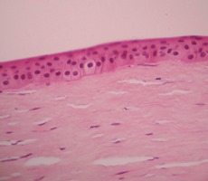
The average corneal thickness greater area ectasia was 344.83 microns and the average was 38.06 microns epithelium. In LASIK cases, the average thickness of the flap and was 162.29 microns 181.34 microns residual stromal bed. The most frequent alterations were atrophy and epithelial hyperplasia (62%), partial loss of Bowman layer (42%) and endothelial (56%) decrease. (Photomicrograph 1).
Discussion
Alterations in subsequent layers of the cornea as a decrease in the stromal bed and endothelial loss, are risk factors for corneal ectasia, which we support with our results; we add alterations in the previous layers.
Conclusions
The findings of the previous layers of the cornea are histopathological factors that may contribute to the formation of corneal ectasia.